
This site is intended for US Healthcare Professionals Only.

Inflammatory bowel disease (IBD) is a chronic disease of the intestinal tract, encompassing both ulcerative colitis (UC) and Crohn’s disease (CD).1,3-5 IBD is marked by inflammation and fibrosis that can contribute to progressive tissue damage and impaired bowel function.3,5-7

In UC, serious complications may include colonic perforation and severe refractory bleeding.8

In CD, fibrosis can contribute to progressive bowel wall thickening and stricture formation, which can result in luminal narrowing and obstruction of the bowel lumen and muscle hypertrophy. Fibrostenosis can lead to a penetrating or stricturing disease course, which impairs quality of life and often requires surgical intervention.9
Maintaining sustained clinical remission has been a challenge for many adult patients with IBD.10

Expanding the Understanding of IBD as an Immuno-Fibrotic Disease3,5,6
Immuno-fibrosis is the process by which inflammation and fibroblast activation drive disease.3,5-7
Immuno-fibrotic diseases are chronic progressive conditions marked by immune dysregulation, inflammation, and fibroblast activation. The impact of inflammation and fibrosis can vary by disease, stage, and patient.3-7


For patients with ulcerative colitis (UC) or Crohn’s disease (CD), both inflammation and fibrosis may impact disease progression, and contribute to long-term complications over time.1-3
In UC, fibrotic structural damage of the colon may contribute to dysmotility, anorectal dysfunction, rectal urgency and incontinence.3 Disease involvement beyond the mucosa may lead to structural damage, and could include submucosal fibrosis and colonic strictures.1
A so-called “lead pipe colon” has also been described in chronic UC, referring to a shortened, stiffened colon on radiological or endoscopic imaging, which may reflect the degree of fibrosis or muscularis mucosae thickening within the bowel wall.11
In CD, chronic intestinal inflammation can lead to complications including fibrotic strictures, fistulas, and abscess formation.3,12 Intestinal fibrosis also presents with strictures and obstructive symptoms, which often require surgical interventions.3,12
Inflammatory Pathways and Fibrotic Mechanisms May Establish a Self-Perpetuating Inflammatory and Fibrotic Loop2,3,5,7
Preclinical studies suggest dysregulation of the immune system can create a cycle in which inflammation induces fibrosis, and the resulting tissue damage exacerbates the immune activation.7,13,14,a

Barrier Dysfunction
Loss of barrier integrity permits increased microbial infiltration, triggering inflammation and an immune response.13
Immune Response
Immune responses can lead to fibroblast activation and release of pro-inflammatory cytokines.9,13
Chronic Inflammation & Fibroblast Activation
Fibroblast activation can lead to excessive ECM deposition.3 Together, fibroblast activation, chronic inflammation, and excessive ECM deposition can contribute to bowel wall thickening, stricture formation, bowel obstruction, and persistent immune dysregulation.2,3,13
Immune Dysregulation
Persistent immune dysregulation can reinforce inflammatory and fibrotic pathways, and may establish self-sustaining immuno-fibrotic loops.7,13
aSupport for this information is based upon preclinical in vitro and animal studies.14

The Immuno-Fibrotic Impact on Inflammatory Bowel Disease Progression3,5,6
Emerging research suggests that immuno-fibrosisb may be integrally involved in IBD progression and could underlie many complications in patients with both CD and UC.2,3,5,6 And while fibrogenesis can be triggered by inflammation, it is also suggested to be an active and self-perpetuating process that may progress in the absence of ongoing inflammatory activity.9,c
bImmuno-fibrosis is the process by which inflammation and fibroblast activation drive disease.3,5-7
cSupport for this information is based upon preclinical in vitro and animal studies.3

Why Fibrosis Matters in IBD
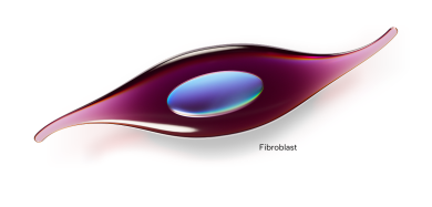
Persistent immune activation can trigger fibroblast activity and excessive ECM deposition—a process of impaired tissue remodeling with clinical implications that include bowel wall thickening, stricture formation, and obstruction.3,15
CD can progress to intestinal obstruction/stenosis, fistula, or abscess, requiring surgical intervention.16
In a retrospective study of newly diagnosed CD patients in the United States between 2017 and 2019, the percentage of patients experiencing CD progression—including intestinal obstruction/stenosis, fistula, intestinal abscess, or CD-related surgery—was 25.2% (1714/6804) over a mean (SD) follow-up of 2.3 years (0.70). 26.6% (444/1668) of newly diagnosed patients experienced CD progression by 3 years.16
Study Design: A retrospective cohort study using an administrative claims and electronic health record database (Optum® Market Clarity) of patients newly diagnosed with Crohn’s disease (CD) in the United States. The primary objective was to characterize CD progression in patients without
CD-related complications. CD progression was defined as the occurrence of intestinal obstruction/stenosis, fistula, or intestinal abscess or certain CD-related surgeries. Patients were selected between January 1, 2016, and June 30, 2019, and followed through June 30, 2020. The study included 6804 patients with newly diagnosed CD and no CD-related complications prior to diagnosis, followed for a mean (SD) of 2.3 (0.70) years. Overall, 25.2% (1714/6804) of patients experienced CD progression during the total follow-up period, with the following CD progression over time: 19.3% (1183/6117) by 6 months, 21.6% (1188/5503) by 1 year, 24.6% (953/3875) by 2 years and 26.6% (444/1668) by 3 years.16
UC can progress to colectomy for medically refractory disease.17
In a retrospective study of adult patients with IBD in the United States, patients newly diagnosed with UC between 2007–2023 were found to have a 5-year cumulative risk of colectomy of 5.7%17
(95% CI, 4.8%–6.6%)
Study Design: A retrospective cohort study using an administrative claims database (OptumLabs Data Warehouse) of adults (18–64 years) newly diagnosed with inflammatory bowel disease in the United States between January 1, 2007, and December 31, 2023. The study included 3893 adults with newly diagnosed ulcerative colitis (UC), and participants were followed over a mean of 4.2 ± 3.0 years.17


A study of colectomy specimens collected in 2007 from 89 UC patients, primarily with extensive colitis and without colonic strictures, found some degree of submucosal fibrosis in 100% of specimens with active inflammation and/or chronic mucosal injury.11
Submucosal fibrosis was almost exclusively detected in areas affected by active inflammation and/or chronic mucosal injury.11
Study Design: The study was conducted in 2018 to characterize the presence, anatomical location, and degree of fibrosis in UC and its association with clinical parameters. Specimens from a total of 89 consecutive UC patients who underwent total colectomy or proctocolectomy at the Cleveland Clinic in 2007 were included; patients with strictures were excluded. The median disease duration at time of colectomy was 62.8 months, and 94.4% of patients had extensive colitis and 91% had refractory disease. 706 individual tissue cross-sections were taken across the 89 colectomy specimens and stained with haematoxylin and eosin and Masson trichrome; a subset of 80 sections was additionally stained with Sirius red and independently reviewed by 3 gastrointestinal pathologists. Degree of inflammation was graded using the validated Geboes score and separated based on histologic features of chronic mucosal injury and active inflammation. Features of chronic mucosal injury and active inflammation were detected in at least one segment from every subject. Submucosal fibrosis was categorized as none, 1%–25%, 26%–50%, 51%–75%, or 76%–100% of the submucosa affected, corresponding to a fibrosis score of 0 to 4.11
Expanding Our Understanding of the Pathways That Contribute to IBD Progression
A growing body of evidence points to the contribution of both inflammatory and fibrotic pathways in IBD progression.3,5,6
Understanding Immuno-Fibrotic Pathways in IBD
IBD progression potentially involves multiple immune-mediated pathways that contribute to inflammation and fibrosis.7 A shift toward pro-inflammatory signalling—including elevated IL-1β, IL-23, IL-17A and IL-17F, alongside IL-6 trans-signalling, which has been linked to chronic intestinal inflammation—has been observed in IBD.18 Furthermore, the cytokines TGF-β, IL-1β, and IL-6 are suggested to be involved in the process of fibrogenesis.3,5,9
The Emerging Science of TL1A

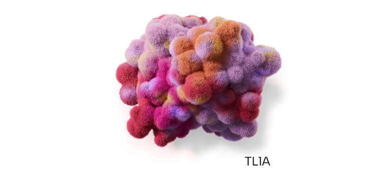
TL1A Is an Amplifier of Immuno-Fibrotic Mechanisms2,b
TL1A may act as a key amplifier within an immuno-fibrotic loop, involved in both inflammatory pathways and fibrotic mechanisms, and contributing to immune dysregulation, chronic inflammation, and fibrosis.2,3,19,20
TL1A, through signaling via the DR3 receptor, may act as an amplifier of innate and adaptive immune responses, and can promote the production of pro-inflammatory cytokines including TNF-α, IL-6, IL-13, and IL-17, while acting in synergy with IL-12 and IL-23 to enhance Th1 and Th17 pathways.2,19,20,b
In addition to its inflammatory effects, TL1A may be associated with a fibrotic response that can contribute to tissue remodeling and fibrotic progression.3,19,b
bSupport for the proposed role of TL1A:DR3 signaling is based upon preclinical in vitro and animal studies.2
Abbreviations
DR3, death receptor 3; ECM, extracellular matrix; IL-1β, interleukin-1 beta; IL-6, interleukin-6; IL-12, interleukin-12; IL-13, interleukin-13; IL-17, interleukin-17; IL-23, interleukin-23; TGF-β, transforming growth factor-beta; Th1, T helper type 1 cell; TL1A, tumor necrosis factor–like ligand 1A; TNF-α, tumor necrosis factor-alpha.

Recognizing Inflammation & Fibrosis as Key Contributors to IBD Progression May Help Move IBD Understanding Forward1-3
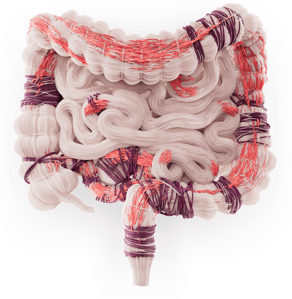
Merck is committed to advancing the science behind immuno-fibrotic diseases, and we encourage continued exploration of immuno-fibrotic pathways to deepen our understanding of IBD progression.1-3
References
- Eggermont E, Gecse K, Krugliak Cleveland N, et al. Ulcerative colitis: moving beyond the mucosal dogma. Lancet Gastroenterol Hepatol. 2026;11(2):163-174. doi:10.1016/S2468-1253(25)00263-8
- Bamias G, Menghini P, Pizarro TT, Cominelli F. Targeting TL1A and DR3: the new frontier of anti-cytokine therapy in IBD. Gut. 2025;74(4):652-668. doi:10.1136/gutjnl-2024-332504
- Rieder F, Mukherjee PK, Massey WJ, Wang Y, Fiocchi C. Fibrosis in IBD: from pathogenesis to therapeutic targets. Gut. 2024;73(5):854-866. doi:10.1136/gutjnl-2023-329963
- D’Amico F, Magro F, Siegmund, et al. Disease clearance as a new outcome in ulcerative colitis: a systematic review and expert consensus. Inflamm Bowel Dis. 2024;30(6):1009-1017. doi:10.1093/ibd/izad159
- Mignini I, Blasi V, Termite F, et al. Fibrostenosing Crohn’s disease: pathogenetic mechanisms and new therapeutic horizons. Int J Mol Sci. 2024;25(12):6326. doi:10.3390/ijms25126326
- Porter RJ, Kalla R, Ho GT. Ulcerative colitis: recent advances in the understanding of disease pathogenesis. F1000Res. 2020;9(F1000 Faculty Rev):294. doi:10.12688/f1000research.20805.1
- Żurawek M, Ziółkowska-Suchanek I, Iżykowska K. Fibrosis in immune-mediated and autoimmune disorders. J Clin Med. 2025;14(18):6636. doi:10.3390/jcm14186636
- Rubin DT, Ananthakrishnan AN, Siegel CA, Barnes EL, Long MD. ACG clinical guideline update: ulcerative colitis in adults. Am J Gastroenterol. 2025;120(6):1187-1224. doi:10.14309/ajg.0000000000003463
- D’Alessio S, Ungaro F, Noviello D, Lovisa S, Peyrin-Biroulet L, Danese S. Revisiting fibrosis in inflammatory bowel disease: the gut thickens. Nat Rev Gastroenterol Hepatol. 2022;19(3):169-184. doi:10.1038/s41575-021-00543-0
- Barberio B, Gracie DJ, Black CJ, Ford AC. Maintenance of clinical remission with biologics and small molecules in inflammatory bowel disease according to trial design: meta-analysis. Dig Liver Dis. 2024;56(1):7-14. doi:10.1016/j.dld.2023.06.009
- Gordon IO, Agrawal N, Willis E, et al. Fibrosis in ulcerative colitis is directly linked to severity and chronicity of mucosal inflammation. Aliment Pharmacol Ther. 2018;47(7):922-939. doi:10.1111/apt.14526
- Lichtenstein GR, Loftus EV, Afzali A, et al. ACG clinical guideline: management of Crohn’s disease in adults. Am J Gastroenterol. 2025;120(6):1225-1264. doi:10.14309/ajg.0000000000003465
- Saha S, Müller D, Clark AG. Mechanosensory feedback loops during chronic inflammation. Front Cell Dev Biol. 2023;11:1225677. doi:10.3389/fcell.2023.1225677
- Li Y, Xu F, Fang Y, et al. Inflammation-fibrosis interplay in inflammatory bowel disease: mechanisms, progression, and therapeutic strategies. Front Pharmacol. 2025;16:1530797. doi:10.3389/fphar.2025.1530797
- Kalafateli M, Tourkochristou E, Tsounis EP, Aggeletoupoulou I, Triantos C. New insights into the pathogenesis of intestinal fibrosis in inflammatory bowel disease: focusing on intestinal smooth muscle cells. Inflamm Bowel Dis. 2025;31(2):579-592. doi:10.1093/ibd/izae292
- Fan Y, Zhang L, Omidakhsh N, et al. Progression of Crohn’s Disease in Newly Diagnosed Patients: Results from an Observational Study Using US Claims Data. Dig Dis Sci. 2024 Nov;69(11):4167-4177. doi:10.1007/s10620-024-08591-7. Epub 2024 Oct 22. PMID: 39438411; PMCID: PMC11567996.
- Ahuja D, Park SK, Qi Y, et al. Treatment and Outcomes of Crohn’s Disease and Ulcerative Colitis in Newly Diagnosed Adults in the United States, 2007 to 2023. Clin Gastroenterol Hepatol. 2026 Apr 15:S1542-3565(26)00280-6. doi:10.1016/j.cgh.2026.04.006. Epub ahead of print. PMID: 41997334.
- Neurath MF. Strategies for targeting cytokines in inflammatory bowel disease. Nat Rev Immunol. 2024;24(8):559-576. doi:10.1038/s41577-024-01008-6
- Tettoni E, Gabbiadini R, Dal Buono A, et al. TL1A as a target in inflammatory bowel disease: exploring mechanisms and therapeutic potential. Int J Mol Sci. 2025;26(11):5017. doi:10.3390/ijms26115017
- Lusetti F, Bezzio C, De Bernardi A, Dota M, Manes G, Saibeni S. The TL1A inhibitors in IBD: what’s in the pot? Expert Rev Gastroenterol Hepatol. 2025;19(1):15-25. doi:10.1080/17474124.2025.2450795